Home >> Atomic-resolution Holography Electron Microscope >> Electron Microscope – How It Works
As most people know, optical microscopes are built based on expanding the principle of the magnifying glass, which magnifies objects using light and a lens. These instruments make it possible to see microscopic worlds by magnifying their visualization several hundreds or thousands of times that of the original size. However, there is a limit to the level of fineness (resolution) that is visible using light. It is not possible to observe objects that are smaller than the wavelength of visible light.
On the other hand, electron microscopes use "electron beams," which have wavelengths much shorter than that of light. These apparatuses emit an electron beam toward the object to be investigated, detect the electrons which pass through, are reflected from or emitted from the object, and create a picture. The brighter and finer the electron beam, the higher the level of observation of the object’s internal details including atomic arrangement.

Optical Microscope
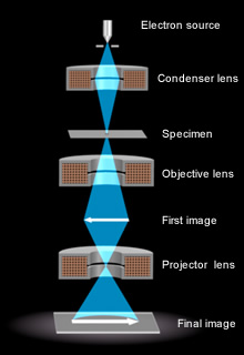
Electron Microscope
Resolution, or more precisely point resolution, refers to the shortest distance in which two points can be recognized as two points. The resolution limit of the optical microscope is determined by the wavelength of visible light; that is, light that can be seen by the human eye. The wavelength of visible light is 400-700 nanometers (nm).The resolution is calculated to be approximately 100 nm. On the other hand, the wavelength of electrons is less than 1/100,000 of that of visible light, or 1 picometer (pm), which is 0.001 nm. Therefore, theoretically, the resolution of electron microscopes can be less than several picometers. However, the resolution obtainable for an electron microscope is restricted to approximately 100 pm by lens aberrations.
1. A convex lens converges the incident parallel light beams. Optical microscopes enlarge images utilizing this convex lens function.
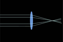
2. Electron microscopes, on the other hand, use electrons to enlarge images, employing an electron lens.
The electron lens uses a magnetic field generated by an electrical current in a coil to converge the electrons.

3. Now, how are the electrons being converged in the magnetic field generated by the coil?

4. To make the explanation easy to understand, let us expand the coil.

5. When electrical current is passed through the coil…
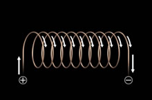
6. A magnetic field is generated.
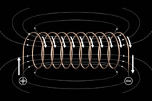
7. Put electrons in the magnetic field; then they travel parallel to the optical axis.
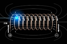
8. These electrons are influenced by the magnetic field

9. In this figure, the direction of the magnetic field is downward. The electrons are moving toward the back; this means that the electrical current is towards the front.

10. Let B the magnetic field and I the electrical current, then electrons are subject to the force F according to the Fleming’s left-hand rule.

11. This means that electrons are circling around the optical axis. and

12. moving toward the back.

13. When the electrons rotate, they are now subjected to the magnetic field parallel to the optical axis.

14. Since the direction of this force is toward the optical axis…,

15. the electrons move along the optical axis, spiraling toward it.

16. As a result, the electron path inside the coil is bent towards the optical axis.

17. Since the magnetic field becomes weaker, when an electron gets closer tothe optical axis, the bending of the electron path becomes smaller ....

18. As a result of this process, all electrons converge at a single point.

19. This is the lens function of the magnetic field generated by the coil.

20. In this way, it becomes possible to enlarge images by electrons in place of light. This mechanism is called an electron lens.

21. Actually, electron lenses…

22. are comprised of doughnut-shaped coils in iron casings.

23. The electron microscope lens system is made up of several electron lenses.

24. In this way, electron microscopes can observe much smaller objects that cannot be seen by optical microscopes.

